Breathing, speaking, walking, laughing, making decisions – it all starts in the head.
Our brain determines perceptions, actions, ideas and emotions, and even our character. Genetic predisposition plays as much of a role as our own experiences and the influences of the environment and people around us. Information enters our brain via the senses, such as sight, touch, hearing or taste. It’s only there that a unique individual image of the world is created. At any given point in time, countless conscious and unconscious processes are running in the human brain. The brain itself changes as a result.
Major advancements in microscopy and other imaging processes are showing with increasing precision how our brain works. But this extraordinarily complex organ continues to pose many questions for scientists. Findings in brain research are not only important in medicine, they also have effects on social areas such as education, parenting and legal practice.
Frontal lobe
The entire front part of the cortex is called the frontal lobe. It controls conscious movement, especially speed, direction and strength. Many scientists also locate the higher cognitive functions of humans here and refer to the frontal lobe as the “carrier of culture”. The frontmost area of the frontal lobe is responsible for attention, deliberation, decision and planning – and it is also considered to be where the personality is located.
Temporal lobe
The best-known function of the temporal lobe is hearing. The auditory centres occupy almost the entire surface of the temporal lobe. Language and music probably require such a high “processing power”. But the temporal lobe is also needed for many other things, for smell, speech, understanding, image recognition and forming memories.
Hippocampus
The hippocampus is a “curled up” section of the cortex and a central element of the limbic system. It’s important for storing knowledge and experiences – anyone without this won’t be able to remember new things. The hippocampus is one of the few areas of the brain in which new nerve cells are made throughout life.
Limbic system
The limbic system is a group of brain areas that are of great importance for the production and processing of emotions, and for memory processes. The most important ones are the hippocampus, the amygdala, the gyrus cinguli and the gyrus parahippocampalis. These brain areas are closely linked with each other. The limbic system controls our emotions and our sexuality, as well as evaluating the importance of information about the outside world.
Hypothalamus
The hypothalamus controls important functions such as reproduction, nutrition, temperature regulation and time measurement. It’s a superordinate centre of the autonomous nervous system that controls unconscious processes, for instance breathing or heartbeat. The rear section of the hypothalamus belongs to the limbic system.
Pituitary gland
The pituitary gland (hypophysis) is only around the size of a pea – but it’s vital. As the “master gland” it controls the body’s endocrine system. It is controlled by the hypothalamus and secretes hormones into the blood. This regulates body functions like growth and reproduction, as well as metabolism.
Cerebellum
The cerebellum is located at the back of the skull. From a perspective of evolutionary history it’s an ancient part of the brain. The connections between the nerve cells are far less complex here than they are in the cerebrum. The cerebellum coordinates motor skills such as posture and walking, but also complex motion sequences such as writing. Despite its small size the cerebellum contains four times as many cells as all the rest of the brain put together.
Brain stem
The brain stem is directly connected to the spinal cord and might be described as the brain’s “technology centre”. No larger than your thumb, the brain stem controls and regulates unconscious vital processes in the body including circulation, breathing and sleep. It is the oldest part of the brain in terms of developmental history. For this reason the differences between humans and animals are comparatively small here.
Communication is everything
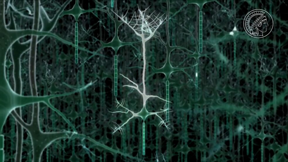
Our brain is a complex network of billions of nerve cells – or neurons – in perpetual communication with each other. Connections are constantly being created or separated, strengthened or weakened. This is also the basis on which we are able to learn and forget. The nerve cells receive electrical impulses via the dendrites and send these to the neuron body. From there they are conducted via the axon to other nerve cells. Transmission from one cell to another happens at the synapses. At this point the electrical impulse is translated into a chemical impulse. There are nerve cells in the brain that receive signals from up to 10,000 other nerve cells, and neurons that pass signals on to thousands of others.
")
 ©")
 © Max Planck Society
The nerve cells in the brain are arranged in layers. These layers and their many connections are the basis for rapid processing of information.
© Max Planck Society
The nerve cells in the brain are arranged in layers. These layers and their many connections are the basis for rapid processing of information.


 ©")


